Skip to main navigation Skip to main content Skip to page footer

Your reconstruction project

You are ready to start your individual reconstruction project? Here is how it works.

Step 1: Image Data

The reconstruction will be carried out on maximal 300 individual slice images (more on request). They can be provided in two ways:

  • OpenSlide-compatible scanner format (see compatible formats here, it is recommended to be verified first with 1-2 sample images)

  • Provide the individualized slice image set of the object to be reconstructed (preferably .png or .tif image format), for a discount of 10% off!

Step 2: First evaluation

In a first step, if we are not sure that the data will be suitable for your reconstruction goal, we will do a test reconstruction based on a set of 20-50 images in lower resolution. This will help us to see if there are any significant problems within the data that might prevent a good quality reconstruction. 

Step 3: Full reconstruction

If the tests were successfull, we will proceed to do the full reconstruction, meaning a full reconstruction using all relevant images in the previously discussed resolution. Typically, this might include an overview reconstruction using the entire slide images, plus a detail reconstruction of the target structures. Details will be discussed based on your specific goals. 

Step 4: Visualization

Together with you, we will find the optimal visualization of your target structures. Get images to visually enhance insights of your research, make others take a second look! The standard reconstruction project contains visualizations based on different gray-level versions of the final data set.

Special requests

You have something special in mind? 

  • Further individual renderings of segmented structures
  • 3-D Segmentation images (volume files)
  • Segmented structures (e.g. exported as .stl)
  • Tell us about your idea and we will find a way to make it work!

Scope of Delivery

What we will deliver

The transformed data set belongs to you, of course. In total, we will deliver the follwing elements:

  • Transformed RGB image data set (.jpg, .tif, ...) with aligned slices
  • 3-D Color Volume Image
  • Gray-Level Volume Image for direct Volume Rendering
  • Volume Rendering images with detail visualizations according requests
  • In case of special options: depending on option, e.g. stl models, additional segmentation images etc.
  • Last but not least: The customer purchases the exclusive, unrestricted right of use for the delivered 2D and 3D images!

Important notes

While we always strive for the best quality reconstruction of your data, please be aware of the following:

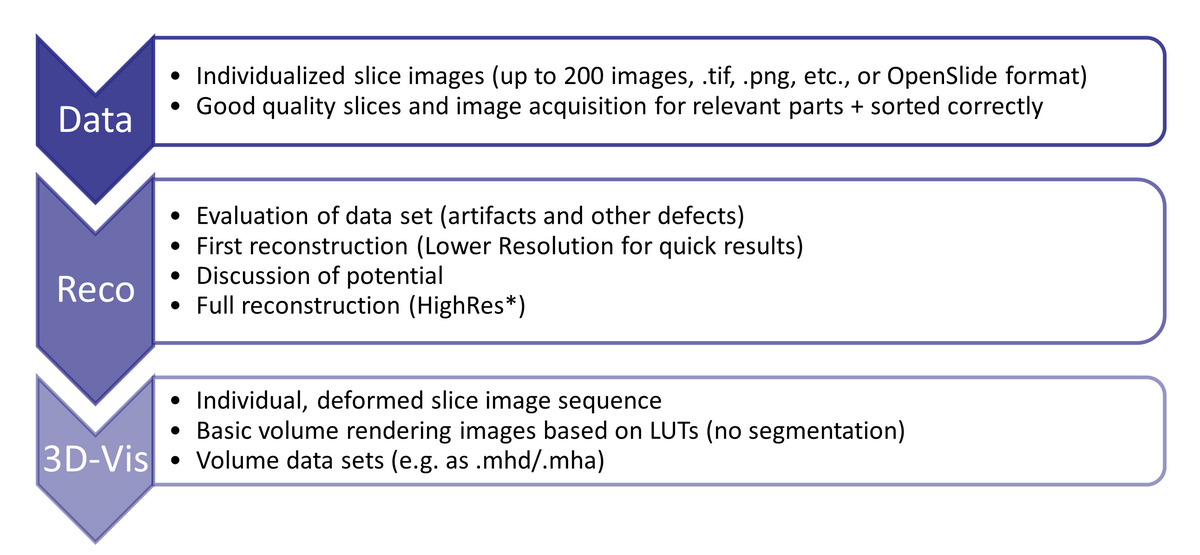
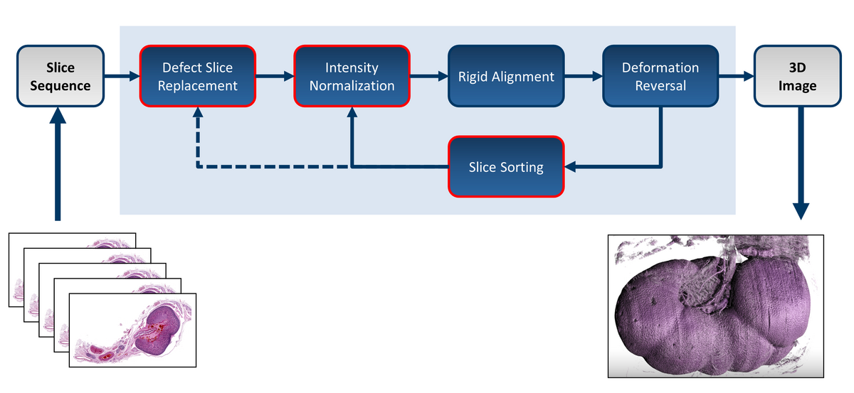
  • The reconstruction quality of histology slice sequences depends mainly on the quality of the slice image data set, and will be adversely affected by artifacts like holes, tears, foldings, missing elements, large distortions, wrong slice order or severe variances in staining, as well as artifacts from scanning (e.g., incorrect, or variable white balance or out-of-focus areas). After a first inspection of the data set, critical defects will be discussed with the customer. 

  • There is unfortunately no guarantee the desired reconstruction result can be achieved with the data set at hand.

  • Due to the extensive data size, a reconstruction and visualization of the full data set in original resolution is typically not possible due to computational limitations. The best visual appearance is achieved with images where the pixel resolution matches the slice thickness. This is typically achieved with a magnification of 2x – 5x. The optimal image resolution for the individual reconstructions depends on the target structures of interest and will be discussed and specified during the project. 

We recommend to refer to our section about specimen preparation, and a discussion about your goals prior to the project.